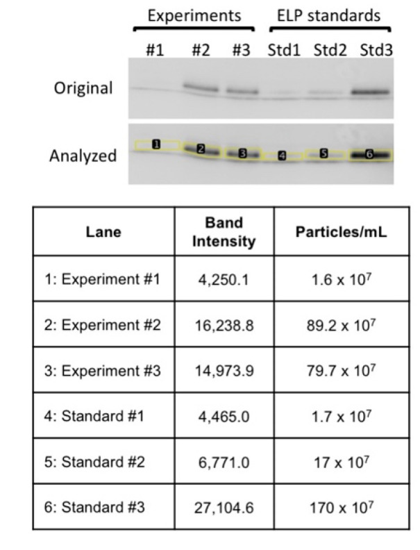
Figure A2.
Western blot-based densitometric analysis for residual LP-A96 in purified EV samples. Purified EVs from three independent experiments (Experiments #1~3, lane 1~3) were blotted for ELPs using anti-ELP antibody AK1 [30] to detect and quantify residual LP-A96 co-purified with exosomes. To determine this, firstly, particle concentration of pure LP-A96 was determined (particle number/mL) using nanoparticle tracking analyzer. Secondly, serial dilutions of LP-A96 were prepared and their particle concentration was correlated to the band intensity in Western blot (ELP Standard #1~3, lane 4~6). Lastly, the standard curve generated based on this correlation was used to extrapolate the LP-A96 particle amount co-purified in EV samples after LP-A96 treatment (lane 1~3). This calculation was used to reflect the raw increase in exosomes in Figure 3B.

