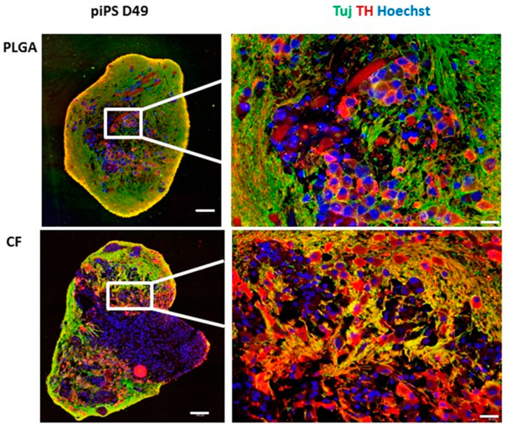
Figure 6.
Immunofluorescent characteristics of the organoids. piPSC PLGA and piPSC CF at day 49 (D49) were collected, fixed in 4% PFA, and then embedded in paraffin blocks and stained with neuron-specific class III β-tubulin (TUJ1), tyrosine hydroxylase (TH), the characteristic marker of mDA neurons, and nuclear marker Hoechst 33342. White scale bar 100 µm (whole organoid, left side of the panel) or 20 µm (fragment of organoid, right side of the panel).

