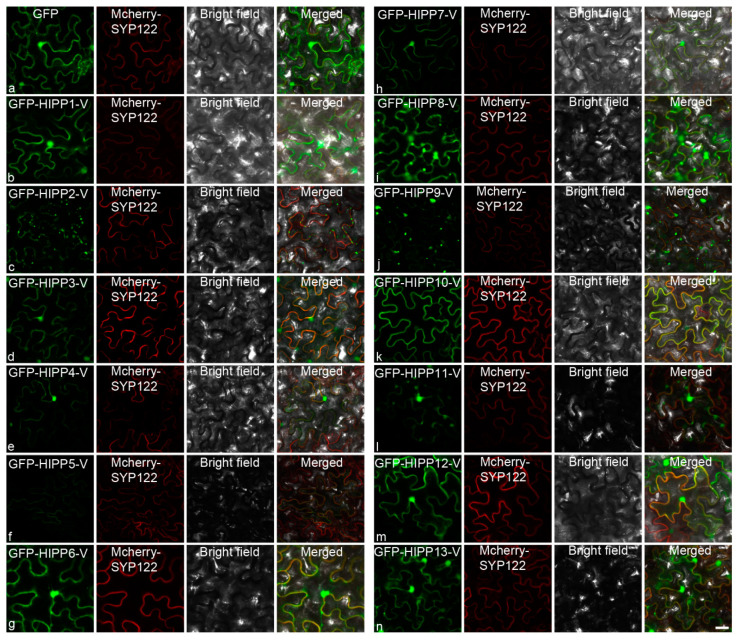
Figure 5.
Subcellular localization of HIPPs in the epidermal cells of Nicotiana benthamiana: subcellular localization of GFP (a), GFP-HIPP1-V (b), GFP-HIPP2-V (c), GFP-HIPP3-V (d), GFP-HIPP4-V (e), GFP-HIPP5-V (f), GFP-HIPP6-V (g), GFP-HIPP7-V (h), GFP-HIPP8-V (i), GFP-HIPP9-V (j), GFP-HIPP10-V (k), GFP-HIPP11-V (l), GFP-HIPP12-V (m), and GFP-HIPP13-V (n). GFP was used as the control. The localization of mCherry-SYP122 is shown in red, and the localization of GFP and its fusion proteins are shown in green. Scale bar = 10 µm.

