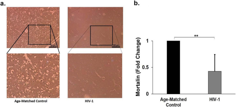
Fig. 9.
Endogenous mortalin is severely reduced in autopsy samples of HIV-1 positive adult brain sections. a) Representative immunohistochemical image of frontal cortex region of adult human brain sections showing expression of mortain (dark brown cells). Section stained in right panel is from a HIV-1 positive individual and left panel is an age matched control, lower panels are the magnified views of indicated regions (n=3). b) Bar graph representd the fold change of mortalin in frontal cortex region of controls and HIV-1 infected individuals. The data represents from three age-matched controls and HIV-1-infected brain sections. Data represented as mean ± Standard Deviation. **P<0.005 as compared with control

