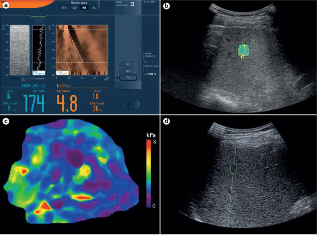
Figure 1 |. Example images from elastographic techniques.
Four methods have been investigated for the assessment of liver fibrosis in patients with NAFLD. a | Vibration-controlled transient elastography (VCTE). The screen (captured directly from a Fibroscan machine; Echosens, France) shows a limited ultrasonic window with a reconstructed view of the wavefront (in brown) that passes through the liver from the handheld probes (M or XL). b | Shear-wave elastography (SWE). SWE is performed in the defined region of interest (ROI). The liver stiffness readout, in m/s, is provided by the software. c | Magnetic resonance elastography (MRE). MRE provides liver stiffness values, in kPa, across the hepatic parenchyma from an average of multiple ROIs. d | Acoustic radiation force impulse (ARFI). This panel is a view of a standard liver ultrasonography image with ARFI performed in the defined ROI (green square). The readout, in m/s, is provided by the software. Image for VCTE (part a) courtesy of Echosens, France. Images for SWE (part b), MRE (part c) and ARFI (part d) were all taken from the same patient at the NAFLD Research Center of the University of California at San Diego, USA.

