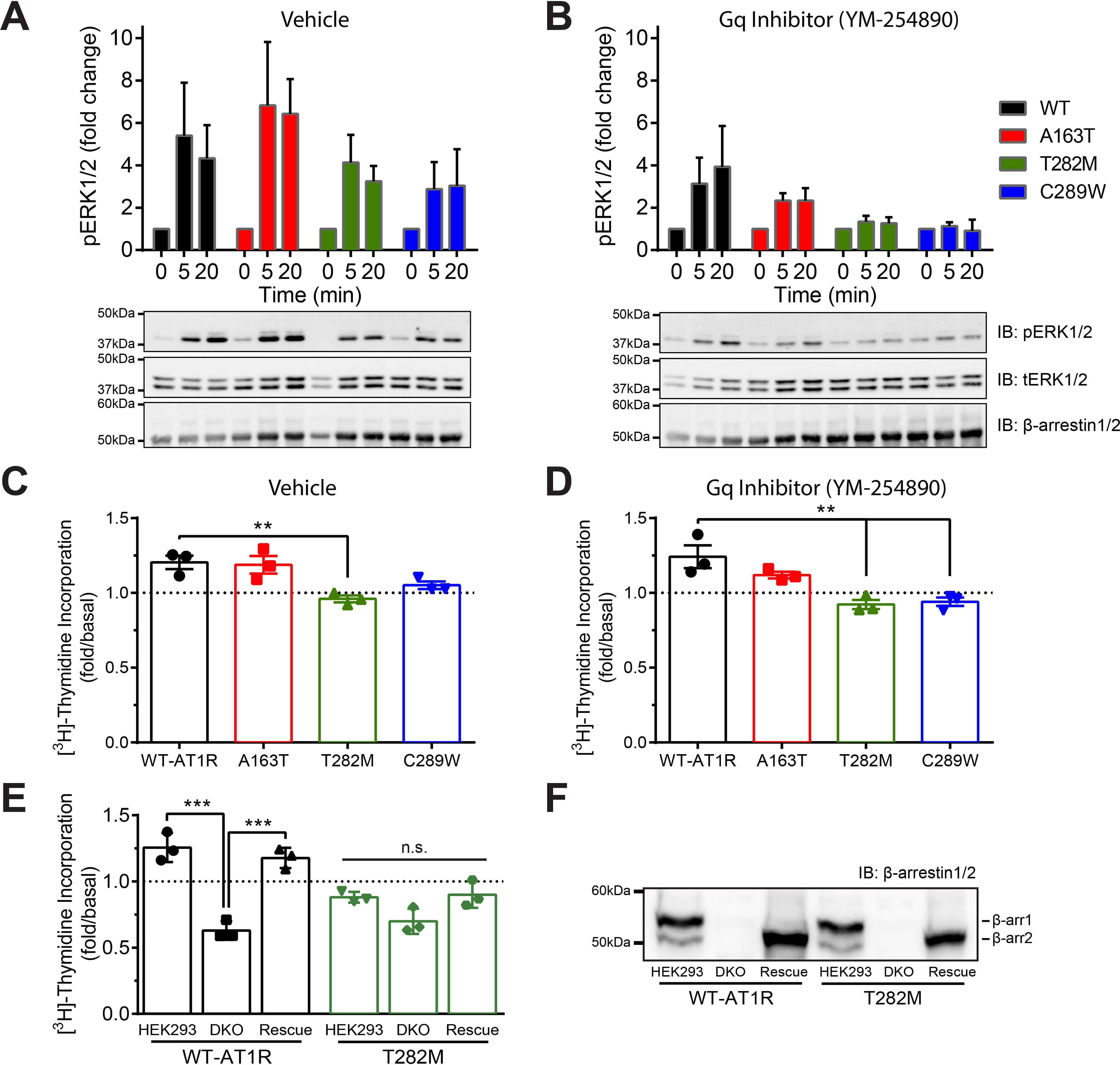
Figure 6.

Role of Gαq/11 and β-arrestin in ERK1/2 activation and proliferation mediated by WT and mutant AT1R. A and B, HEK293 cells were transfected with WT or mutant AT1R and β-arrestin2 and pretreated with vehicle (A) or selective Gαq/11 inhibitor YM-254890 (B) to dissect G protein– versus β-arrestin–mediated ERK1/2 activation. Cells were stimulated with 1 μm AngII for 0, 5, or 20 min and then lysed and analyzed via Western blotting with antibodies specific for phospho-ERK, total-ERK, and β-arrestin1/2. Quantifications are represented as mean ± S.D. from four to six independent experiments. C and D, HEK293 cells were transfected with WT or mutant AT1R and β-arrestin2 and pretreated with vehicle (C) or YM-254890 (D). Cells were stimulated with or without 1 μm AngII for 24 h in presence of [3H]thymidine and then lysed to measure incorporated radioactivity. Data are represented as mean ± S.D. of duplicates from three independent experiments, and one-way ANOVA followed by Dunnett's multiple comparison tests were performed where ** = p < 0.01, n = 3. E and F, HEK293 and HEK293/DKO cells were transfected with WT or mutant AT1R, along with additional β-arrestin2 (rescue). Cells were stimulated with vehicle or 1 μm AngII for 24 h in presence of [3H]thymidine and then lysed to measure incorporated radioactivity. For F, cells from E were lysed and analyzed via Western blotting with antibodies specific for β-arrestin1/2. Data are represented as mean ± S.D. of duplicates from three independent experiments, and one-way ANOVA followed by Tukey's multiple comparison tests were performed for each receptor where *** = p < 0.001, n = 3.
