Figure 7.

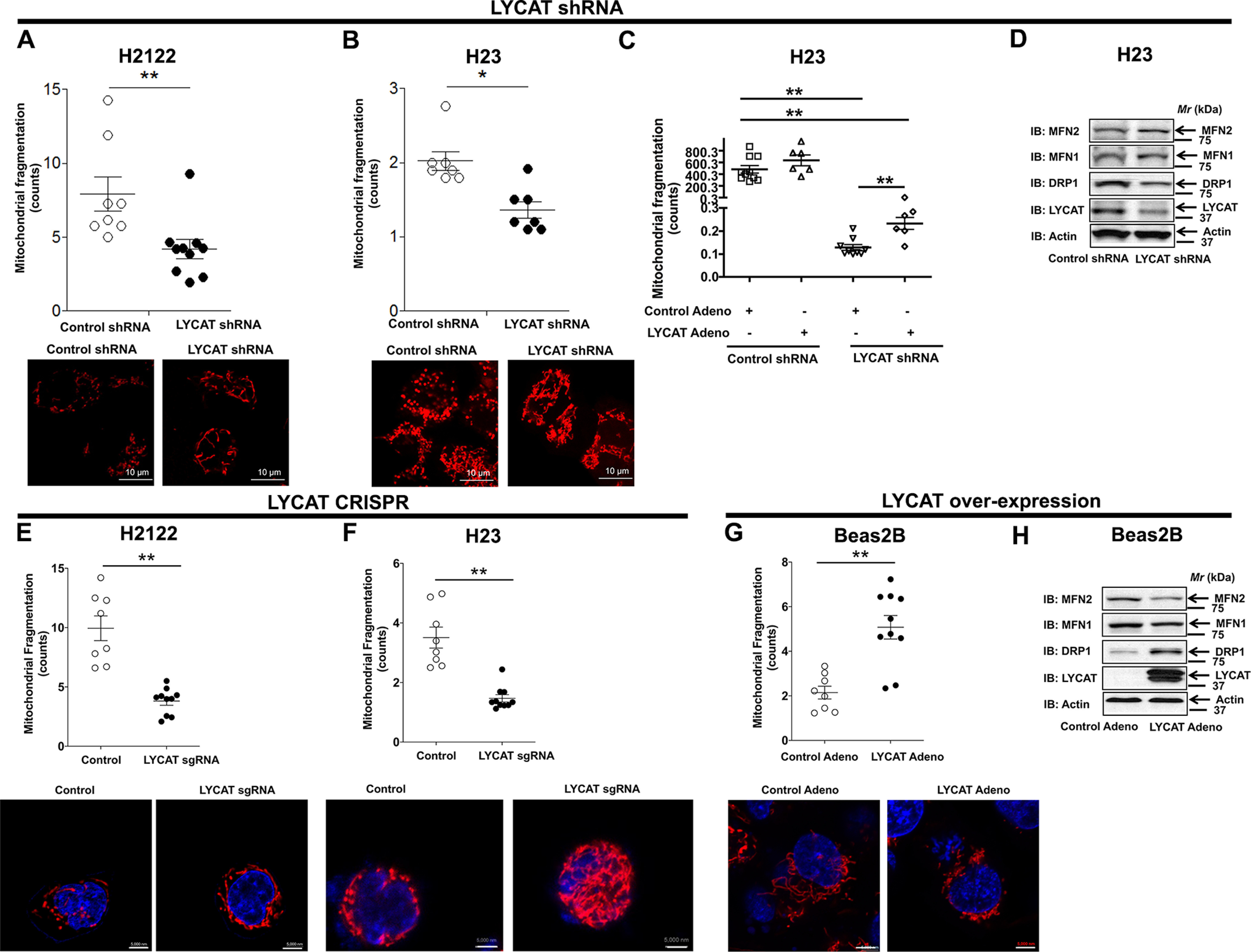
LYCAT knockdown inhibits mitochondrial fragmentation. A and B, H2122 (A) and H23 (B) cells stably expressing either control or LYCAT shRNAs were stained with MitoTracker Red fluorescence dye, and mitochondrial fragmentation was recorded by confocal microscopy. Scale bar, 10 μm. *, p < 0.05; **, p< 0.01 versus parental control. C, restoration of LYCAT expression in H23 LYCAT knockdown clones rescues the effect of LYCAT knockdown on mitochondrial fragmentation, as determined by MitoTracker staining and confocal microscopy. **, p < 0.01 versus parental control. D, expression of DRP1 that drives mitochondrial fission and MFN1/2, which control mitochondrial outer membrane fusion in CRISPR-Cas9 LYCAT–edited H23 cells. The β-actin blots in D and Fig. 6C are identical because they were from the same experiment and run at the same time, when β-actin immunostaining was performed. E and F, LYCAT–edited H2122 (E) and H23 (F) knockout cells were stained with MitoTracker Red fluorescence dye, and mitochondrial fragmentation was recorded by confocal microscopy. Scale bar, 5000 nm. *, p< 0.05; **, p < 0.01 versus parental control. G, LYCAT-overexpressing Beas2B cells were stained with MitoTracker Red fluorescence dye, and mitochondrial fragmentation was recorded by confocal microscopy. **, p < 0.01 versus control. H, cell lysates of LYCAT-overexpressing Beas2B cells were probed for the expression of the indicated proteins via immunoblotting (IB). Error bars, S.E.
