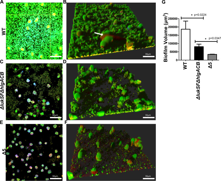
FIG 1.
Leukocidins are required for survival of biofilms exposed to neutrophils. Wild-type USA300 biofilms were incubated with Cell Tracker Blue-labeled primary human neutrophils for 2 h, stained with Syto-9 (viable bacterial cells; green) and ethidium homodimer-1 (DNA of dead mammalian cells; red), and imaged using confocal laser scanning microscopy. (A) Image of a section taken close to the base of the biofilm. (B) Three-dimensional image showing a wild-type USA300 biofilm after a 2-h incubation with primary human neutrophils. The arrow indicates DNA of a dying neutrophil. (C and D) Experiments similar to those for panels A and B, performed with biofilms of a ΔlukSF ΔhlgACB USA300 strain. (E and F) Experiments similar to those for panels A and B, performed with biofilms of a Δ5 USA300 strain, lacking all leukocidin proteins. (G) Volume quantification of WT, ΔlukSF ΔhlgACB, and Δ5 USA300 biofilms after treatment with neutrophils for 2 h. Biofilms were grown in μ-slides and captured at a ×600 magnification. Images represent the majority population phenotype seen in six independent experiments performed in triplicate. Student t tests were performed for pairwise comparisons.

