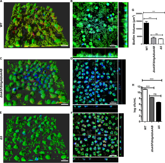
FIG 4.
PVL, HlgAB, and LukAB are required for biofilms to evade neutrophil killing. (A) Neutrophils were incubated with wild type biofilms and imaged after 1 h. Staining of neutrophils and bacteria was done as described for Fig. 1. (B) Representative image of section taken close to the base of wild-type biofilms and imaged as described for panel A. (C and D) Experiments performed similar to those for panel A, with an isogenic ΔlukSF ΔhlgA ΔlukAB mutant of USA300. (E and F) Experiments performed similar to those for panel A, with an isogenic mutant lacking all 5 leukocidins (Δ5 USA300). (G) Biofilm volume quantified after a 1-h incubation of biofilms from the indicated strains with neutrophils. (H) Measurements of total bacteria (CFU per milliliter) isolated from biofilms of indicated strains, after a 1-h incubation with neutrophils. Results are averages from six independent experiments performed in triplicate, with SEM. Enumerations of CFU per milliliter were done independently, using biofilms grown in silicone tubing. Multiple comparisons were done using one-way analysis of variance and Tukey’s post hoc analysis where appropriate. **, P < 0.01; ***, P < 0.001; ns, not significant. Images were taken using Imaris software version X 6.4. MFI calculations were performed using ImageJ.

