FIG 1.
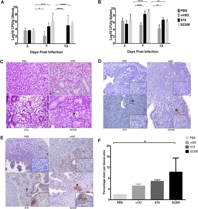
Brucella abortus colonized the uterus and induced endometritis in nonpregnant ICR mice. Mice were inoculated intraperitoneally with 1 × 10⁶ CFU of PBS, B. abortus mutant 2308ΔvirB2 (virB2), live-attenuated vaccine strain S19, or wild-type Brucella abortus S2308. Uterine samples were collected at 3, 7, and 13 days postinfection (dpi), and bacteria were enumerated via culture. (A and B) Bar graphs representing the average (n = 5) with the standard deviation (SD). Statistical differences between the groups were determined using two-way ANOVA and Tukey’s multiple-comparison test. *, P < 0.05; **, P < 0.01; ***, P < 0.001; ****, P < 0.0001. (C) Representative micrographs (hematoxylin and eosin stain) of normal uterus from control and infected nonpregnant mice at 13 dpi, respectively (×60 magnification). Mild to moderate macrophage infiltration of the endometrium and expansion of the endometrial tissue with clear space (edema) was evident. Asterisks represent edema and inflammatory cell infiltration. (D and E) Brucella antigen distribution (D) and positive immunolabelling of macrophages (E) in the uterus of the respective mice at 13 dpi. Asterisks indicate Brucella antigens or Iba-1-positive macrophages, respectively. (F) Percentage stain area in the uterus of nonpregnant mice. Estimation of stain area intensity was performed with Image J analysis. Significance was estimated using one-way ANOVA and Tukey’s multiple-comparison post hoc test. *, P < 0.05.

