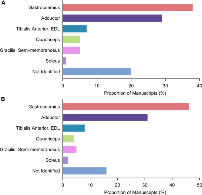
Figure 7.

Breakdown of muscles analyzed in the published literature evaluating postischemia angiogenesis. A, Graph showing the prevalence of the specific muscles used for histological angiogenesis evaluation and quantitation, among all manuscripts analyzed (n=509 manuscripts). B, Graph showing the prevalence of the specific muscles used for histological angiogenesis evaluation, after author-level adjustment to account for multiple manuscripts from the same research group (n=283 manuscripts). EDL indicates extensor digitorum longus.
