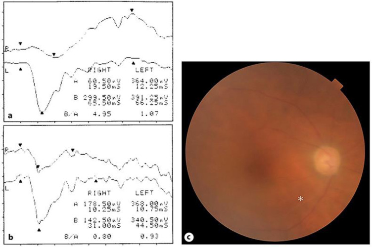
Fig. 3.
a ERG recorded before the cataract surgery. The ERG of the right eye shows a reduced amplitude and prolonged implicit time in the a-wave compared with that of the left eye. b ERG of the right eye 4 months after DSAEK. Improvement is seen in the amplitude and the implicit time due to the improved transparency of the ocular media. c Fundus photograph taken 4 months after DSAEK. Though the image of the fundus is hazy because of the residual corneal stromal edema, the color of the retina looks normal and there are no signs of atrophy in the optic disc, but a ghost vessel can be seen in the lower temporal area (asterisk). DSAEK, Descemet stripping automated endothelial keratoplasty; ERG, electroretinogram.

