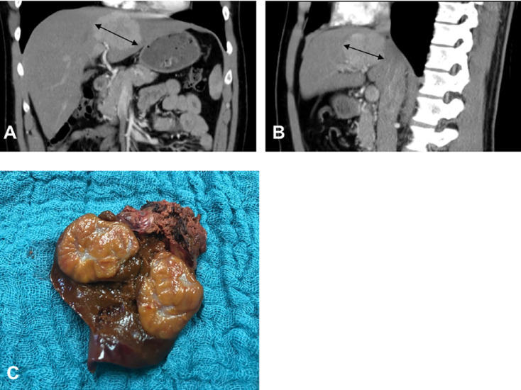
Fig. 3.
A 30-year-old male patient with a focal nodular hyperplasia in liver segment IV. A CT, coronal section, showing focal nodular hyperplasia 7 cm in diameter (bidirectional arrows). B CT, axial section. C The depicted operative specimen indicates atypical liver resection of segment IV with a central scar on the cut surface.

