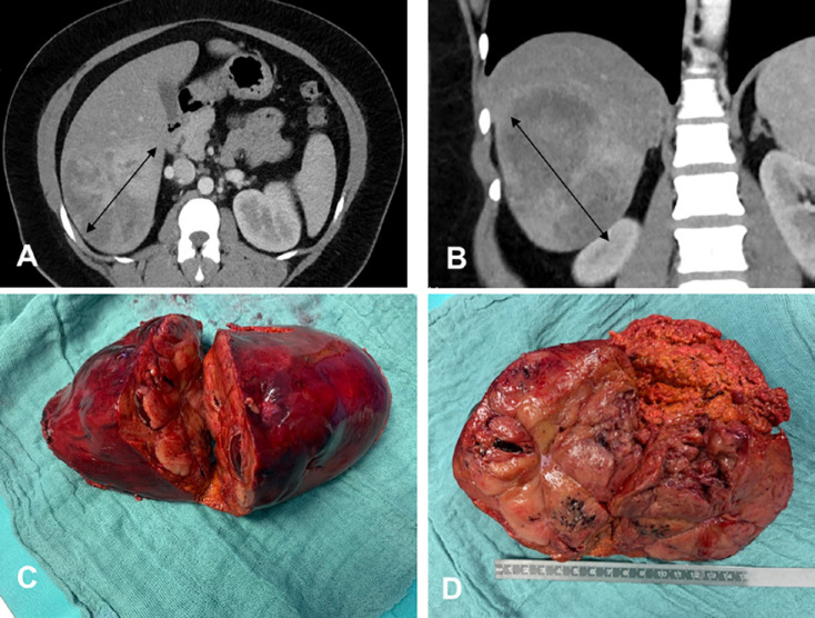
Fig. 5.
A 32-year-old female patient with hepatic adenoma. A The tumor is 10 cm in diameter (bidirectional arrows) in segments VI and VII, coronal section. B CT, axial section. C, D Surgical specimen of an atypical liver resection of segments VI and VII (C) and of normal hepatic parenchyma and hepatic hemangioma visible on the cut surface (D).

