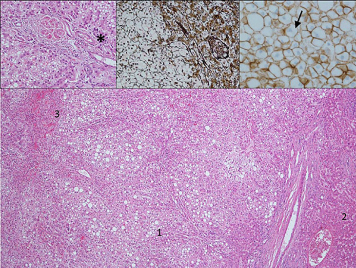
Fig. 6.
Hepatocellular adenoma with fatty changes limited to the lesion (1) but absent in normal liver (2). Hemorrhage is a common feature (3). Insets: Higher magnification (left inset) does not show portal tracts but so-called naked arteries (*) and, in comparison to hepatocellular carcinoma, no nuclear atypia or mitotic activity. Reticulin (middle inset) with Gomori silver staining demonstrates a preserved reticulin framework somewhat slightly reduced in the lesion versus the normal liver. In this case, β-catenin staining (right inset) was negative with only membranous and no nuclear reactivity. Brown, cell membrane; blue, nucleus.

