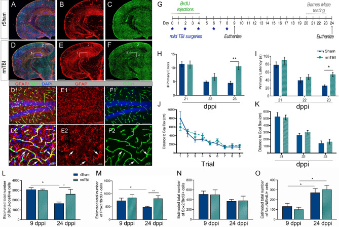
Figure 1.
rmTBI induces GFAP immunoreactivity, learning and memory impairments, and expansion of neuroblast/immature neurons. (A–C) Representative maximum projection z-stack confocal mosaic image of serial section stained for GFAP (red), CD31 (green) and DAPI (blue) in rSham at 24 dppi. (D–E) Representative confocal image at 24 dppi rmTBI showing increased GFAP immunoreactivity in the hippocampus (D1–F1; insert) and cortex (D2–F2; insert). (G) Experimental timeline showing repeated mild TBI surgeries (*blue), daily BrdU injections (green), behavioral testing (grey), and two time-points for tissue collection (arrows, black). The rmTBI mice show an increase in the # primary errors (H) and latency in seconds (s) (I) made during Barnes Maze testing at 23 dppi. No difference was found in the distance to goal box per trial (J) or day (K). The number of BrdU (L) and Prox1/BrdU-positive (M) cells in the DG was increased in rmTBI mice while no change was seen in Sox/BrdU (N) or NeuN/BrdU (O) at 24 dppi compared to rSham. Scale = 1 mm in (A–F); 500 µm in (D1–F1) and 20 µm in (D2–F2). Data are represented as mean ± SEM. Two-way ANOVA for repeated measures with Bonferroni post-hoc. *p < 0.05, and **p < 0.01. n = 10–15 mice per group for behavior and n = 7–12 for stereology.

