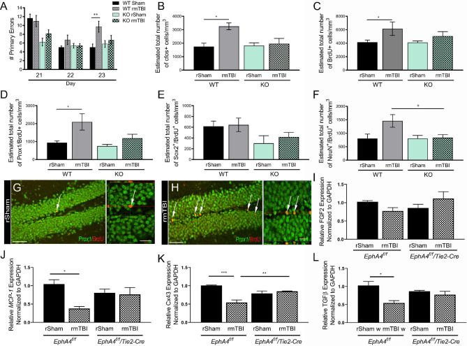
Figure 4.
EphA4f./f/Tie2-Cre mice show attenuation of rmTBI-induced deficits. (A) The number of primary errors in significantly increased in rmTBI EphA4f./f (WT) mice at 23 dppi compared rSham. This effect was not observed in EphA4f./f/Tie2-Cre (KO) mice. Compared to rSham, rmTBI KO mice did not show an increase in the number of cFos (B), BrdU (C) or Prox1/BrdU (D) positive cells in the DG as was seen in WT mice. No difference in the number of Sox2/BrdU (E) or NeuN/BrdU (F) positive cells were found across the groups of mice. (G–H) Representative confocal images of Prox1/BrdU positive cells in the DG at 24 dppi in WT mice. (I–L) Quantified mRNA expression of total DG tissue using qPCR for FGF2, MCP-1, Gja1 (Cx43), and TGFβ, respectively. All genes were normalized to GAPDH then represented relative to WT rSham levels. *p < 0.05; **p < 0.01, ***p = 0.001. Scale = 100 µm in (G,H); 20 µm in inset. n = 5–10 mice per group for stereology and n = 10–16 for behavior. qPCR was performed using biological triplicates.

