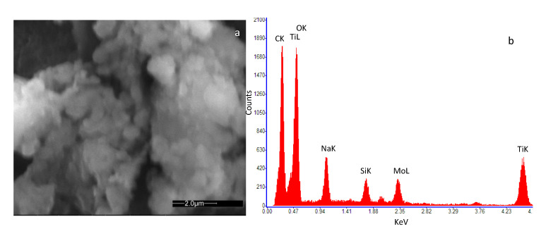
Figure 3.
High-resolution field emission scanning electron microscopy (SEM) image of the deposited film of SWCNTs decorated by TiO2 nanoparticles. The microscopic investigation was performed with a Quanta 400 (FEI) emission scanning electron microscope, operated at 20.0 kV. (a) view of the deposited film; (b) result of the Energy Dispersive Spectroscopy (EDS) on the analyzed sample.

