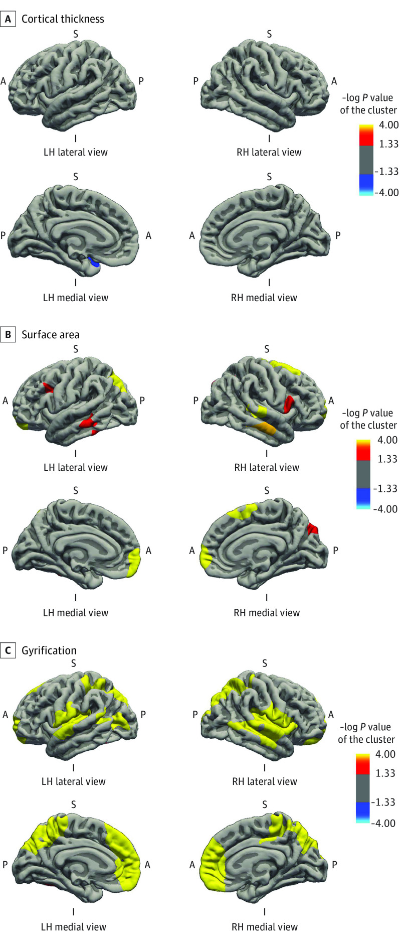
Figure 3. Gestational Age at Birth and Cortical Thickness, Surface Area, and Gyrification in Children Born at Term.
Surface-based analysis was performed for 2706 children with a gestational age at birth ranging from 37.0 weeks to 41.9 weeks. The model was adjusted for child sex, child age at neuroimaging, maternal ethnicity, maternal age at intake, marital status, educational level, psychopathologic conditions during pregnancy, smoking and alcohol use during pregnancy, and family income. Colored clusters represent regions of the brain that were positively (red to yellow) and negatively (dark to light blue) associated with gestational age at birth that survived the clusterwise (Monte Carlo simulation with 5000 iterations) correction for multiple comparisons (P < .001) (eTable 3 in the Supplement). A indicates anterior; I, inferior; L, lateral; LH, left hemisphere; M, medial; P, posterior; RH, right hemisphere; S, superior.

