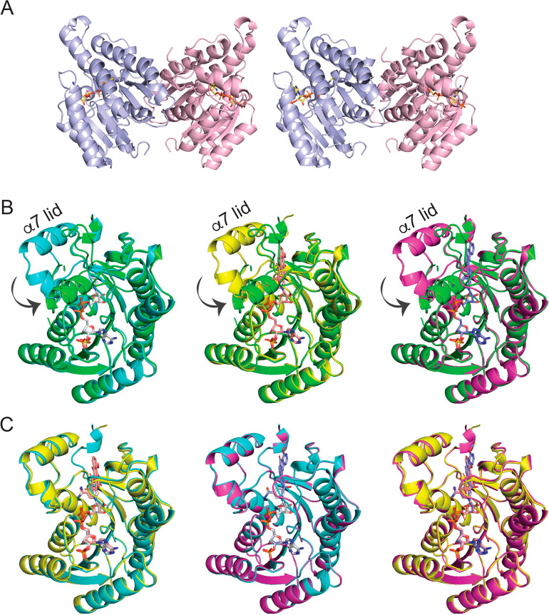
Figure 3.
Dimeric arrangement of LugOII structures and observed conformational changes. (A) Stereoscopic view of NADPH-bound LugOII in dimeric form. (B) Unliganded LugOII (green), LugOII/NADPH (cyan), LugOII/NADPH/4 (yellow), and LugOII/NADPH/5 (magenta) structures are superimposed. α6, α7, and the loop region between them serve as a lid, which turn around 180° and then rotate 90° toward the binding site of 5. (C) Alignment of LugOII/NADPH (cyan), LugOII/NADPH/4 (yellow), and LugOII/NADPH/5 (magenta) structures. NADPH, 4, and 5 are displayed in sticks.

