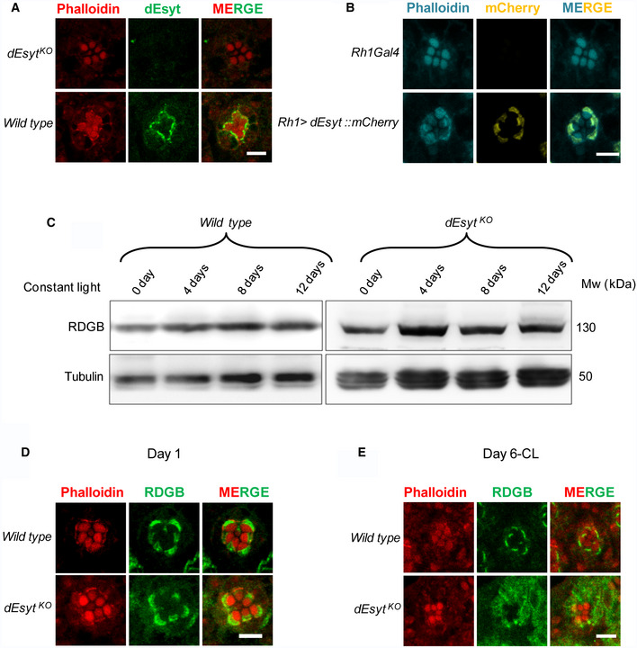
Figure 4. dEsyt localizes to MCS and determines RDGB localization.

-
AConfocal images showing the localization of endogenous dEsyt protein in photoreceptors of one‐day-old dark‐reared flies probed with antibody against dEsyt. Scale bar: 5 μm. dEsyt KO shows no staining when probed with dEsyt antibody.
-
BConfocal images showing the localization of exogenously expressed dEsyt::mCherry protein expressed using the eye‐specific Rh1‐Gal4 in one-day‐old dark‐reared flies. Rh1‐Gal4 is shown as a control. A single ommatidium is shown. Scale bar: 5 μm. Phalloidin marks F‐actin staining and highlights rhabdomeres R1–R7.
-
CWestern blot from the head extracts of wild type and dEsyt KO probed with the antibody against RDGB. Rearing conditions, age of the flies, and genotype is indicated on top of the blot. Tubulin was used as the loading control.
-
D, EConfocal images showing the localization of RDGB in wild‐type and dEsyt KO photoreceptors of flies which are (D) 1‐day-old dark‐reared and (E) 6‐day-old exposed to constant illumination. For (D) and (E) RDGB visualized using an antibody against the endogenous protein. Rhabdomeres are outlined using phalloidin which marks F‐actin. Scale bar: 5 μm.
