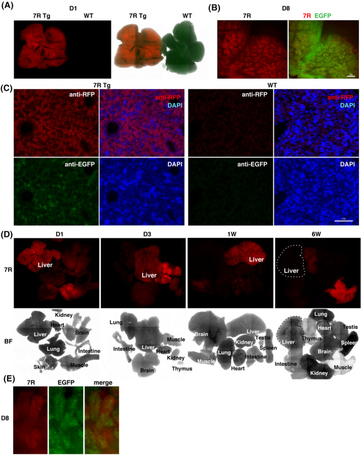
Figure 2.

Parenchymal hepatocytes specifically express RFP in the newborn liver. A, 7R, CYP3A7R transgenic liver; WT, wild‐type liver on day 1, a negative control. B, Transgenic hepatic lobules on day 8. Scale bar, 500 µm. C, Liver sections immunostained for RFP on day 1. Scale bar, 100 µm. D, CYP3A7R mouse organs. 7R, RFP fluorescence signals; BF, bright field. E, Enlarged image of intestinal microvilli on day 1
