
Figure 7.
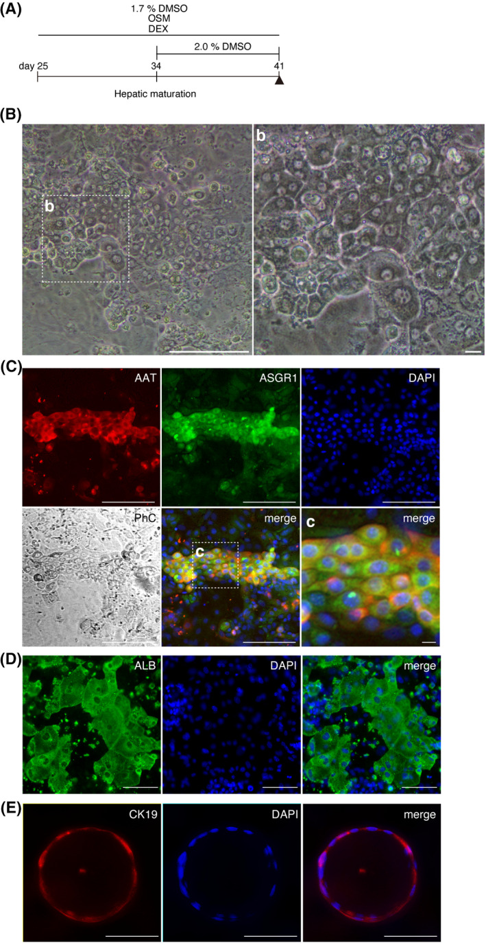
HB‐LCs capable of differentiating into HLCs and bile ducts. A, Maturation culture method (n = 4). The day‐41 cells matured from day‐25 HB‐LCs. B, Phase‐contrast microscope image of the day‐41 cells. Scale bar, 100 μm (left). (b) Enlarged image of the framed area in (B). Scale bar, 10 μm. C, Immunostaining for mature hepatocyte markers, AAT, and ASGR1. Scale bar, 100 μm. (c) Enlarged image of the framed area in (C). Scale bar, 10 μm. D, Immunostaining for ALB. E, Biliary epithelium marker, CK19. Scale bar, 100 μm