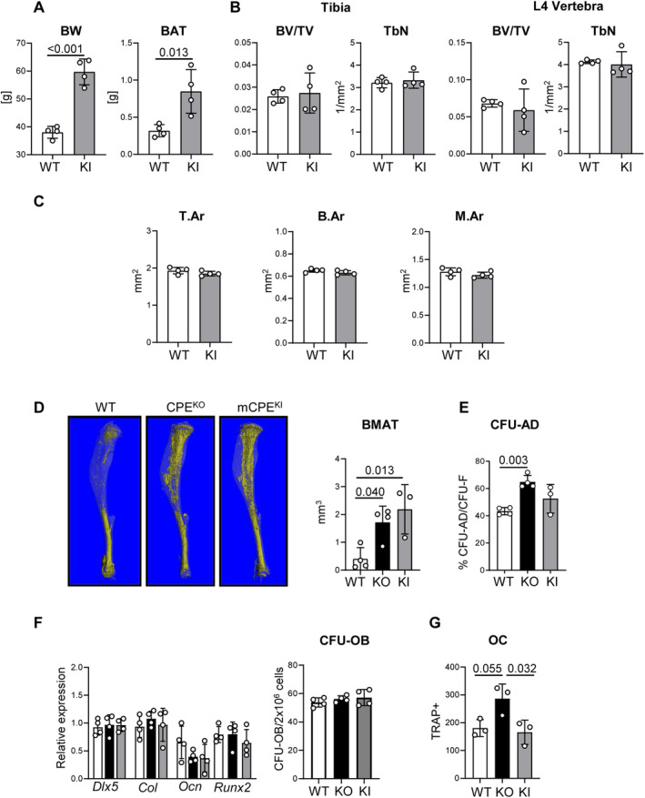
Fig 2.

Mutated carboxypeptidase E knockin (mCPE KI) male mice are obese, but do not display trabecular bone deficit at the age of 40 weeks. (A) Body weight and weights of brown adipose tissue in WT and mCPE KI males. (B) μCT analysis of trabecular bone in proximal tibia and L4 vertebrae. BV/TV = bone volume per tissue volume; TbN = trabecular number. (C) μCT analysis of tibia cortical bone. T.Ar = cortical bone total area; B.Ar = cortical bone area; M.Ar = marrow area in diaphysis. (D) μCT renderings of BMAT stained with osmium tetroxide in a whole tibia and volumetric measurements of bone marrow adipose tissue (BMAT) in proximal tibia of WT, CPE KO, and mCPE KI mice. (E) Bone marrow stromal cell differentiation to adipocytes measured in colony‐forming units for adipocytes (CFU‐AD) assay. (F) Osteoblast‐specific gene markers expression in bone marrow stromal cell and osteoblastic differentiation measured in colony‐forming units for osteoblasts (CFU‐OB) assay. (G) Marrow nonadherent cell differentiation to tartrate‐resistant acid phosphatase‐positive (TRAP+) multinucleated osteoclast‐like cells (OC) in the presence of 50 ng/mL RANKL and 50 ng/mL macrophage colony‐stimulating factor. White bars = WT; black bars = CPE KO; gray bars = CPE KI; n = 4 mice per group.
