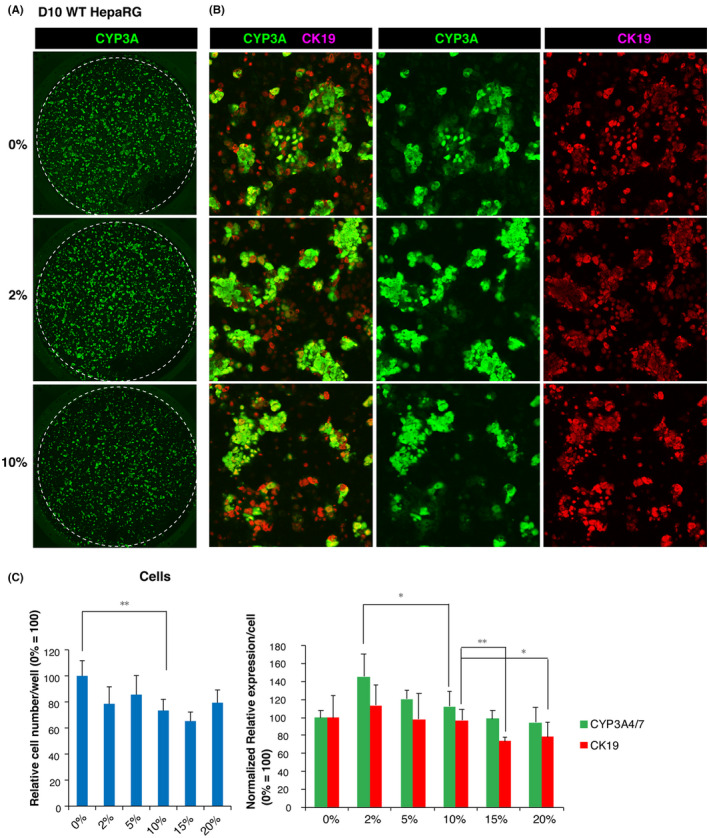
FIGURE 7.

Optimized 2D culture system for the formation of hepatocyte‐like cells (HLCs) from HepaRG cells and biliary epithelium on O plates coated with 2% Cellmatrix Type I‐A. A, Images of CYP3A4‐positive cells (green) and CK19‐positive cells (red) generated by confocal microscopy (upper). An enlarged view of the center of the well is shown in B. C, Semi‐quantitative fluorometric evaluation of HLCs and the biliary epithelium according to the ICC signal intensity for CYP3A4 and CK19, respectively. Cell growth under the indicated conditions was estimated based on the fluorescence intensity of DNA staining with Hoechst 33 258 (cells). Normalized relative values were calculated according to the average value obtained from D10 cells cultured on noncoated O plates taken as 100 (n = 4)
