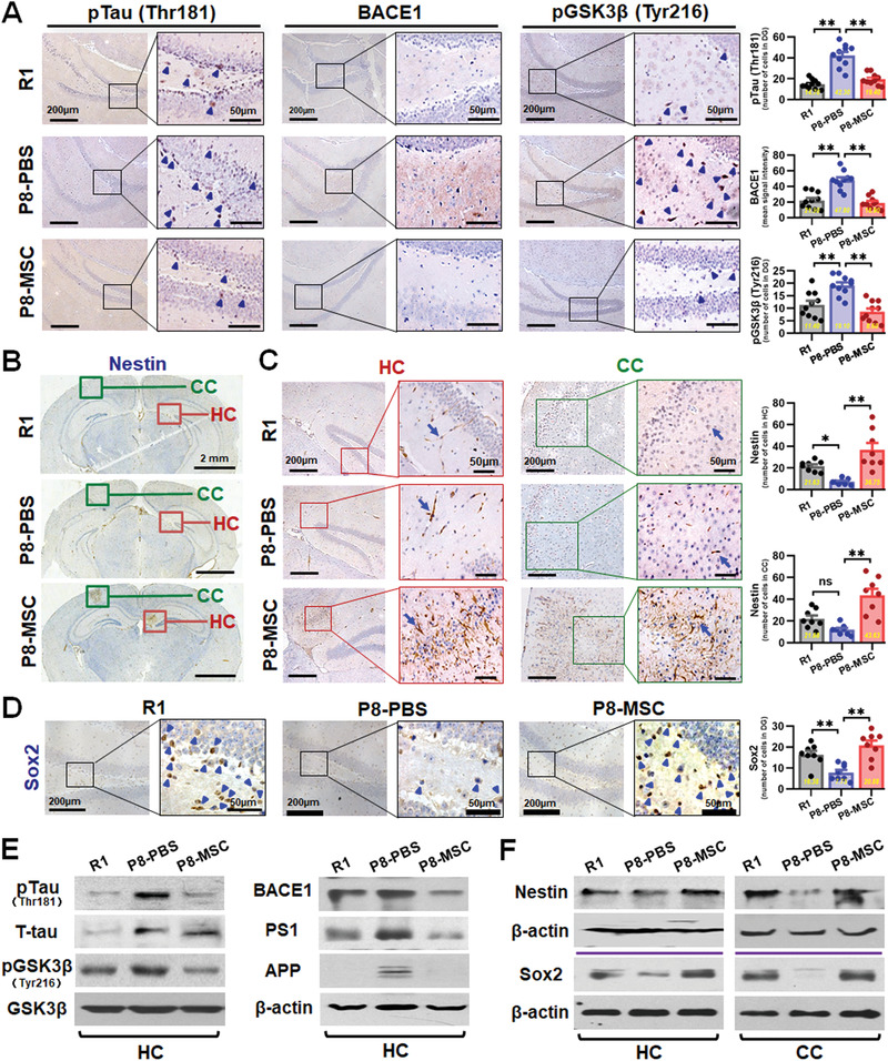
Figure 2.

hUC‐MSCs regulated the expression of AD‐related key proteins and improved neurogenic regeneration in SAMP8 mouse brain. A) Representative images of hippocampal dentate gyrus (DG) showing the expressions of pTau (Thr181), BACE1, and pGSK3β (Tyr216) in R1, P8‐PBS and P8‐MSC mice by immunohistochemical method. Squares in higher‐magnification inserts indicate the protein positive cells with arrowheads‐labeled individual cell. Right: quantification of pTau (Thr181), BACE1, and pGSK3β (Tyr216) in DG region of the R1, P8‐PBS, and P8‐MSC groups. B,C) Immunohistochemical analysis the effects of hUC‐MSCs on the endogenous neurogenesis. B) Representative images illustrated the overall distribution of the Nestin+ stem cells from the coronal hippocampal sections. Green circles and red circles respectively represent the specific region of cortex and hippocampus which Nestin+ stem cells were concentrated and further showed the higher‐magnification in (C). Images and the number statistics mice for proliferative around CA1 region (C, left) and specific region of cortex (C, right). D) Images and the number statistics for proliferative Sox2+ stem cells in DG. E) Western blotting results showing the expressions of pTau (Thr181), pGSK3β (Tyr216), APP, BACE1, and PS1 in the hippocampus of the R1, P8‐PBS, and P8‐MSC groups. F) Western blotting results showing the expressions of Nestin and Sox2 in hippocampus and cortex of the three groups. (n = 8–10 per group; all data shown as mean ± SEM, *P < 0.05, **P < 0.01).
