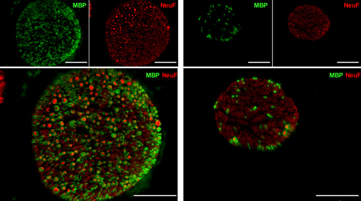
Figure 2.

Representative image of a murine cervical (left panel) and abdominal (right panel) VNs. The nerve fibers (NeuF) are depicted in red, and the myelin (MBP) is illustrated in green. Scale bars are 50 µm. NeuF: neurofilament F, MBP: major basic protein
