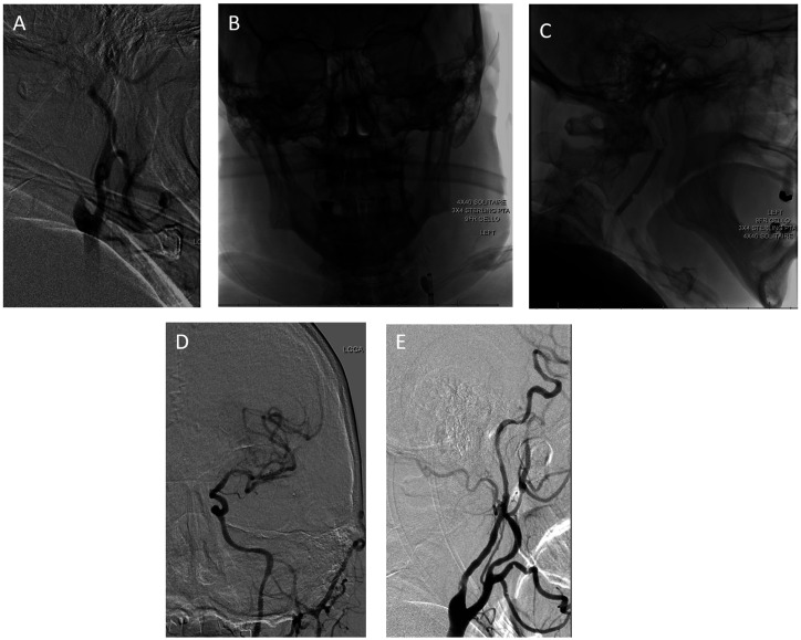
Figure 1.
(a) Lateral view of the left cervical carotid artery demonstrating occlusion at the proximal origin of the internal carotid artery; (b and c) single-frame AP and lateral shots, which demonstrate a solitaire stent retriever deployed in the M1 segment with a Sterling balloon inflated across the cervical carotid occlusion. The cello guide catheter is also inflated during inflation of the cervical balloon to provide some proximal embolic protection; (d) final AP cranial view from the left common carotid artery which demonstrates complete recanalization of the MCA candelabra; (e) final lateral cervical view, which demonstrates recanalization of the origin of the internal carotid artery.

