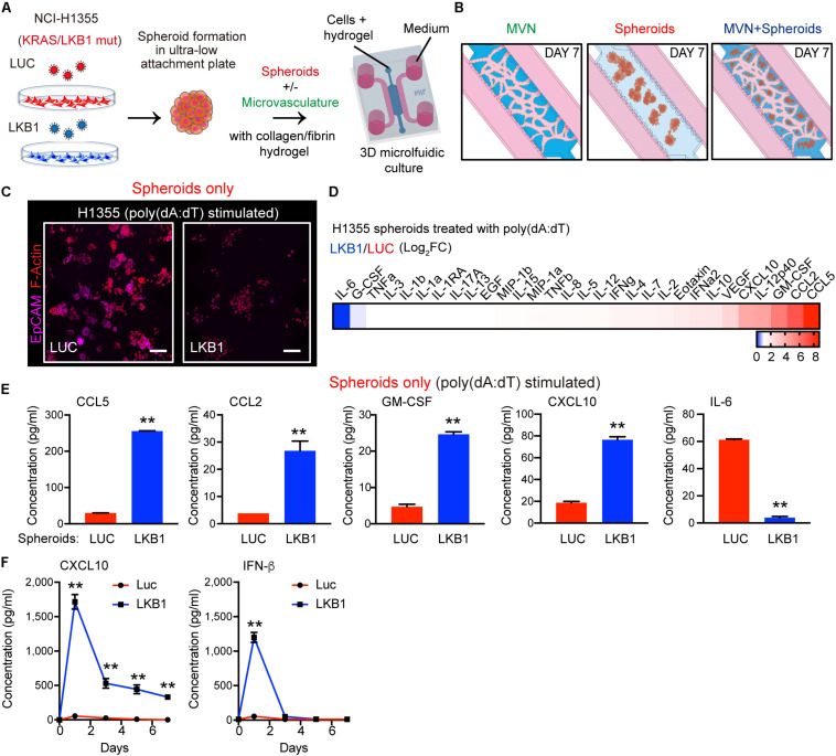
FIGURE 1.
LKB1 reconstitution of 3-D KL spheroids and response to dsDNA in microfluidic culture. (A) Schematic of H1355 tumor spheroids formation and their in vitro dynamic coculture with or without the microvasculature (MVN) in a 3D microfluidic device within a collagen/fibrin hydrogel. (B) Schematic of the dynamic culture of microvasculature only, spheroids only and the combination of spheroids and microvasculature in the microfluidic culture. (C) Confocal image of luciferase (LUC) control expressing (left) and LKB1 reconstituted (right) H1355 spheroids in 3D microfluidic culture after 7 days, pre-stimulated with poly 1 μg/mL poly (dA:dT), immunostained for F-actin (red) and EpCAM (CD326) (violet). Scale bar, 150 μm. (D) Heat map of cytokine profiles in conditioned medium (CM) 7 days from 3D microfluidic culture of H1355 spheroids. CM was collected 7 days after pre-stimulation with 1 μg/mL poly (dA:dT). Values represent log2 fold change of LKB1 reconstituted H1355 spheroids relative to control. (E) Absolute values of cytokine release of human CCL5, CCL2, GM-CSF, CXCL10, and IL-6 produced from 3D microfluidic culture of H1355 LKB1-reconstituted spheroids versus control. (F) ELISA of human CXCL10 and IFN-β over 7 days of 2D culture, treated ± 1 μg/mL poly(dA:dT), (n = 3 biological replicates). CM was collected and refreshed daily. P values were calculated by unpaired two tailed student t-test; **P < 0.01. Data shown as mean values, error bars ± SD.

