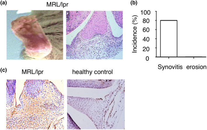
Figure 1.

IgG deposition in arthritic joints of lupus mouse. (a) Representative images of ankle and histopathology of the knee joint in MRL/lpr mice at the age of 30 weeks. (b) Incidence of synovitis and bone erosion in MRL/lpr mice at the age of 30 weeks (n = 8). (c) Representative images of immunohistochemistry staining of IgG in the joints of MRL/lpr mice and C57BL/6 mice at the age of 25 weeks.
