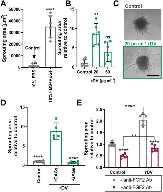
Figure 3.

rDV promotes endothelial cell angiogenic sprouting. HUVEC spheroids were embedded in a fibrin gel and cell sprouting from the spheroid body was analyzed from phase contrast micrographs after 24 h. A) Establishment of an appropriate negative control, demonstrating that little HUVEC sprouting occurred in the presence of 10% FBS, compared to 10% FBS with 5 ng mL−1 VEGF165, making 10% FBS an appropriate negative control (Control). All other experimental conditions contained 10% FBS in addition to the test substance. B) HUVEC sprouting in the presence of 20 and 50 µg mL−1 rDV. Cell sprouting is expressed as fold change relative to Control and **p < 0.01 is relative to Control. C) Representative images of HUVEC spheroid under Control conditions or in the presence of 20 µg mL−1 rDV. Scale bar is 200 µm. D) HUVEC sprouting in the presence of 20 µg mL−1 rDV (+GAGs) or with 20 µg mL−1 rDV with GAG chains removed by enzymatic digestion (‐GAGs). Cell sprouting is expressed as fold change relative to Control and ****p < 0.0001 is relative to rDV+GAGs condition. E) HUVEC sprouting under Control or 20 µg mL−1 rDV in the absence (‐anti‐FGF2 Ab) or presence (+anti‐FGF2 Ab) of the anti‐FGF2 antibody, demonstrating that cell sprouting involves FGF2 signaling. Cell sprouting is expressed as fold change relative to Control and ****p < 0.0001 is relative to the corresponding anti‐FGF2 Ab condition unless indicated otherwise. Data are mean ± SD (n = 5–6).
