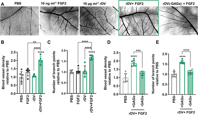
Figure 5.

rDV promotes angiogenesis in vivo by potentiating growth factor signaling via its GAG chains. rDV was added to the CAM at embryonic day 8 (E8) and the effect on the vessel density and the number of branch points was studied at E12. A) Representative dissecting microscope images of the blood vessels in the CAM at E12 following exposure to PBS, FGF2 (10 ng mL−1), rDV (10 µg mL−1), or the two combined (rDV (10 µg mL−1) + FGF2 (10 ng mL−1)). Scale bar is 1 mm. B,C) Blood vessel density (B) and number of branch points (C) in CAM membranes exposed to PBS, FGF2 (10 ng mL−1), rDV (10 µg mL−1), or the two combined (FGF2 (10 ng mL−1)+ rDV (µg mL−1)). D,E) Blood vessel density (D) and number of branch points (E) in CAM membranes exposed to PBS or a combination of rDV and FGF2 (rDV (10 µg mL−1) + FGF2 (10 ng mL−1)) with GAG chains on rDV intact (+GAGs) or removed via enzymatic digestion (−GAGs). Data are expressed as fold change relative to PBS. Data are mean ± SD (n = 5–6). **p < 0.01 and ****p < 0.0001 relative to PBS unless indicated otherwise.
