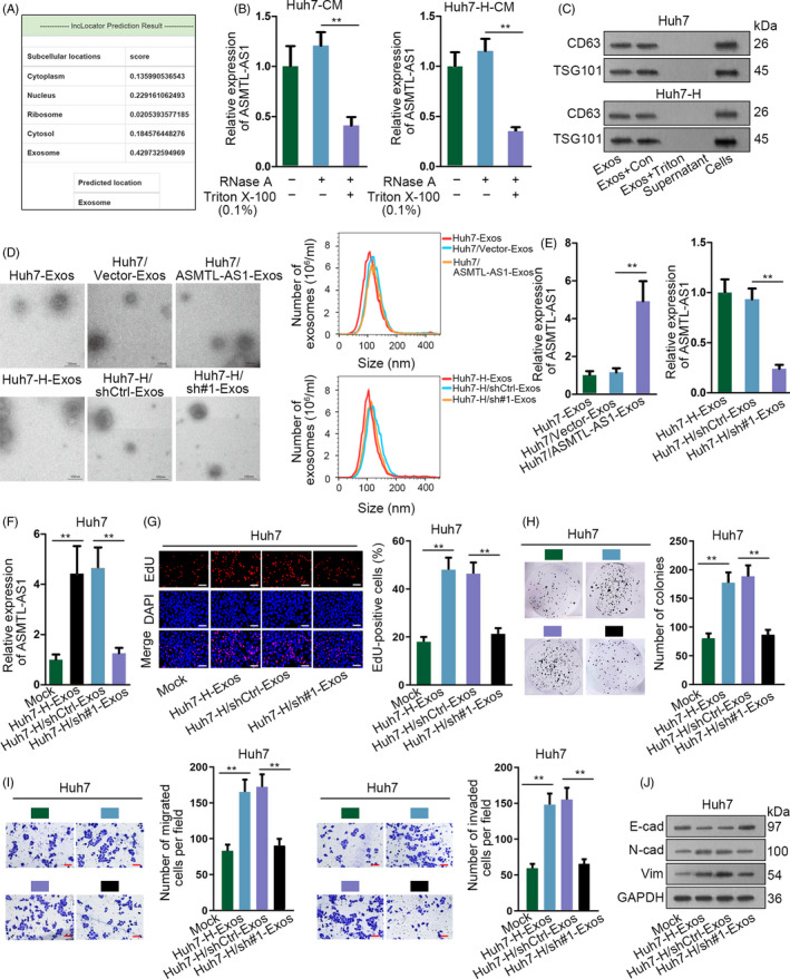
Figure 6.

Exosome‐transferred ASMTL‐AS1 delivered malignancies between HCC cells. A, Online tool lncLocator predicted that ASMTL‐AS1 mainly located in exosomes. B, ASMTL‐AS1 level in Huh7 and Huh7‐H under diverse treatments was estimated by qRT‐PCR. C, Western blot identified exosomes through two markers including CD63 and TSG101. D, Representative images of exosomes from indicated cells were captured by electron microscopy, and the size and number of these exosomes were determined via NanoSight particle tracking analysis. Scale bar = 100 nm. E, The level of ASMTL‐AS1 in above exosomes was analysed through qRT‐PCR. F‐J, Impact of exosomes from control or ASMTL‐AS1‐silenced Huh7‐H cells on the cellular processes of Huh7 cells was estimated by EdU assay (scale bar = 200 μm), colony formation assay, transwell assay (scale bar = 200 μm) and Western blot. **P < .01
