
Figure 5.
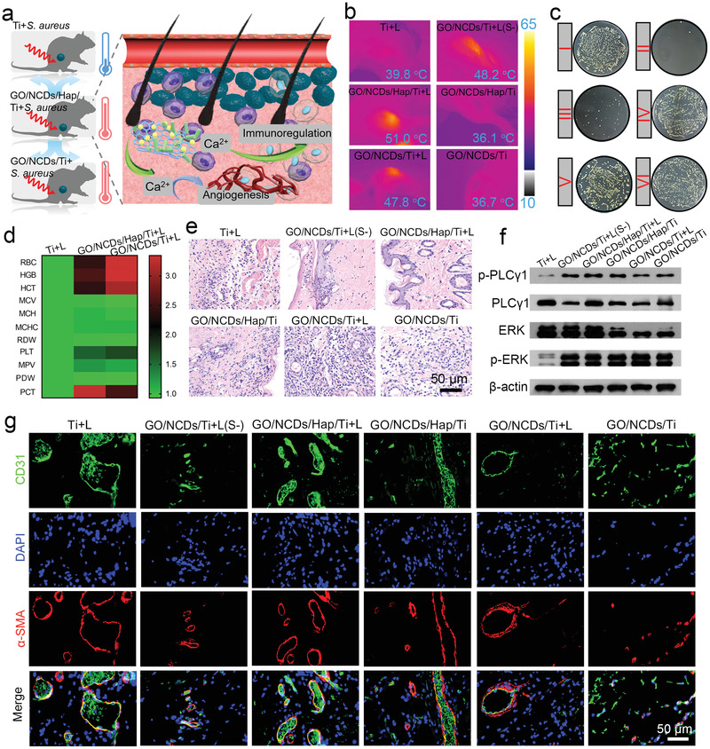
a) The schematic diagram of mild phototherapy through the NIR induced Ca2+ flow and then activated the PLCγ1/ERK and PI3K/P‐AKT pathway for injured vessel repair and inflammation relieve. b) The photothermal images in vivo after NIR irradiation (0.5 W cm−2, 15 min). c) The corresponding spread plates of S. aureus after NIR irradiation with (b). d) The blood routine examination of Ti+L, GO/NCDs/Ti+L, and GO/NCDs/Hap/Ti+L at 1 D treatment. e) The H&E staining of infected tissue near the implant at 1 D treatment. f) The WB analysis regarding to the vascular repair pathway at 3 D treatment. g) The immunofluorescent staining of CD31 (green), DAPI (blue), and α‐SMA (red) at 6 D treatment.