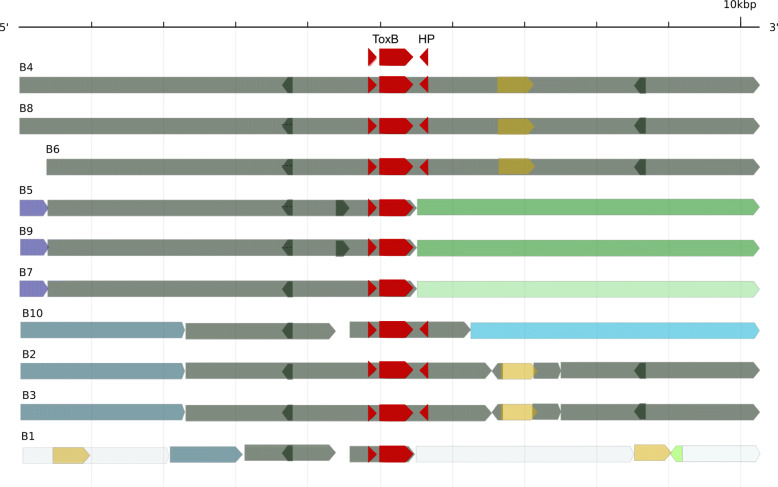
Fig. 6.
DW5 ToxB 10 kb locus region (B1-B10) sequence homology. Regions in common are represented by the same colours relative to the ToxB gene position (red). For example, a small 5′ region is shared by B5, B7 and B9 and is shown in purple. Grey regions are common to all ten loci and gaps are shown in white. The hypothetical protein (HP) is found downstream of ToxB in B2–4, 6, 8 and 10. Regions of similarity are shown between forward sequence (forward arrow) and reverse sequence (reverse arrow). Short regions of similarity contained within a larger region are secondary alignments

