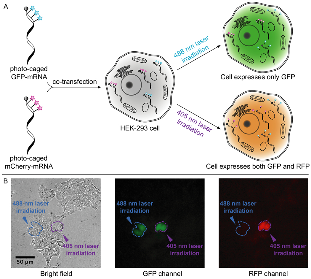
Figure 3. Live cell photo-activation of mRNA translation.

A) HEK-293 cells are co-transfected with caged GFP-mRNA and caged mCherry-mRNA, followed by photo-uncaging with either 488 nm (30 seconds) or 405 nm laser (10 seconds) irradiation. B) Live cell fluorescence imaging. Selected cells (circled in blue) irradiated with 488 nm laser only express GFP, shown as green cells in the GFP channel and dark cells in the RFP channel. Selected cells (circled in purple) irradiated with 405 nm light express both GFP and RFP. Scale bar = 50 μm.
