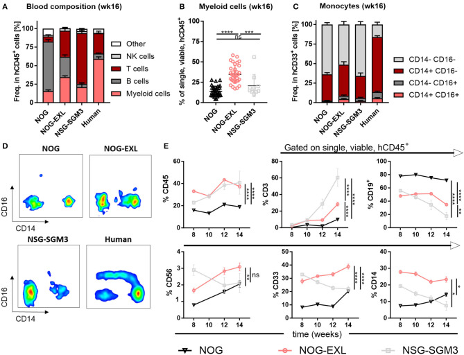
Figure 2.
Peripheral blood analysis demonstrates improved human immune cell reconstitution. (A) Human leukocytes composition (CD45+) in peripheral blood (PB) 16 weeks after reconstitution as percentage, n = 28–40. (B) Comparison of human myeloid cells (CD33+) in human leukocytes (CD45+) as percentage from same mice as in A, one-way ANOVA, Tukey's multiple comparison test. *p < 0.05; ***p < 0.0005; ****p < 0.0001, n = 10–39. (C) Human monocyte composition in total CD33+ in PB 16 weeks after reconstitution or in human PBMCs as percentage, n = 5–40. (D) Representative FACS plots for human monocyte populations (CD16+/−/CD14+/− in CD45+) in PB of humanized mice in week 16 or in human PBMCs. (E) Different immune cell populations (CD45+ in single viable; CD19+, CD3+, CD33+, CD56+, and CD14+ in CD45+) in HIS-NOG, HIS-NOG-EXL and HIS-NSG-SGM3 over time (week 8–14), ordinary 2-way ANOVA, *p < 0.05; **p < 0.001; ***p < 0.0005; ****p < 0.0001, n = 28–121.

