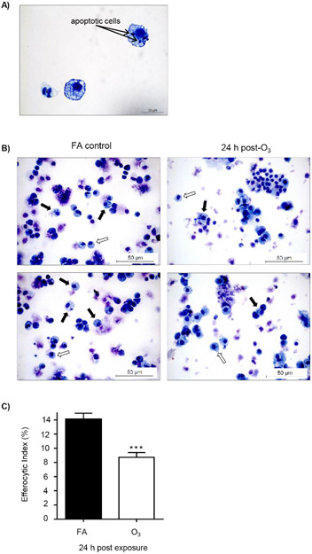
Figure 3: O3 exposure decreases alveolar macrophage efferocytosis.
C57BL/6J female mice were exposed to filtered air (FA) or 1 ppm O3for 3 h. 24 h post-exposure, mice were oropharyngeally instilled with approximately 5 x 10⁶ apoptotic Jurkat T cells. 1.5 h after instillation, bronchoalveolar lavage (BAL) was performed, and the efferocytic index was calculated in BAL macrophages by light microscopy after counting 200 macrophages (n = 11 per group). (A) Representative image of an efferocytic macrophage. (B) Identification of alveolar macrophages (white arrows) and efferocytic macrophage (black arrows) after FA or O3 exposure. (C) Calculation of the efferocytic index after FA or O3 exposure (***p < 0.0001).

