Figure 1.

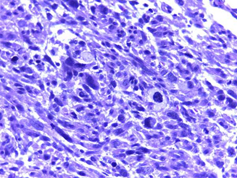
Photomicrograph of haematoxylin and eosin histologic section (×100) shows interlacing fascicles of atypical spindle cells with patchy necrosis, abnormal mitoses and smudge cells.

Photomicrograph of haematoxylin and eosin histologic section (×100) shows interlacing fascicles of atypical spindle cells with patchy necrosis, abnormal mitoses and smudge cells.