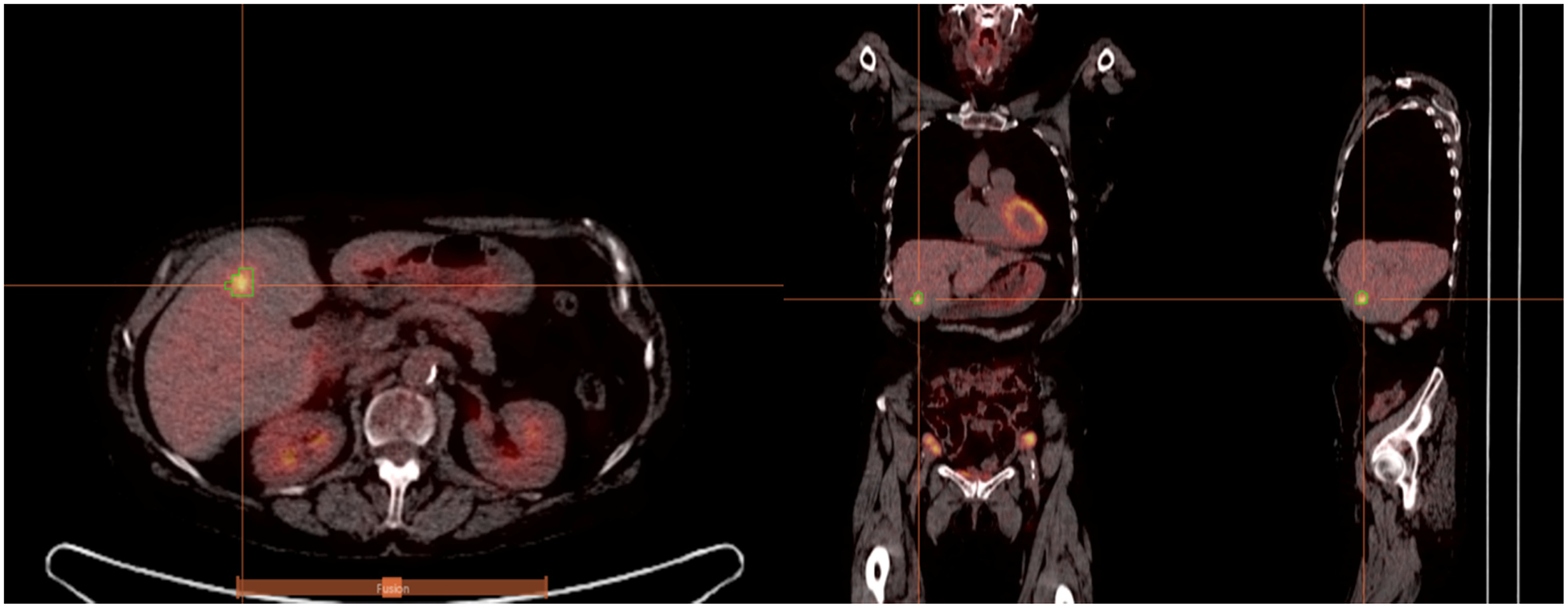
Figure 1.

PET/CT images from a patient with colorectal liver metastasis. Sagittal, coronal, and transaxial slices (left-to-right) are shown. Overlaid on the images is 3D PET-derived segmentation via 40% background-corrected SUVmax thresholding. Tumors were identified based on the PET images, though the fused PET/CT images were used initially to ensure that the tumors were intrahepatic (and not metastases in lung or peritoneum).
