
Figure 2.
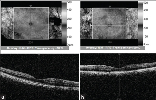
(a) Showing optical coherence tomography of the right eye showing good foveal contour and absence of cystoid edema. (b) Showing left eye good foveal contour, focal loss of inner segment-outer segment line in the nasal parafoveal area and absence of cystoid edema