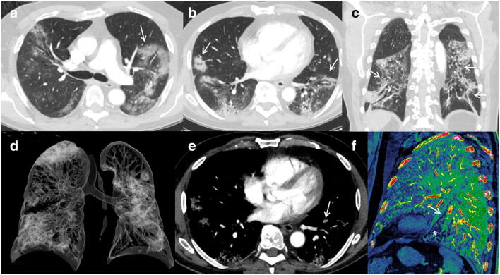
Fig. 2.
Segmental left lower lobe PE over a severe lung involvement. CTPA, with lung window (a, b, c) and volume rendering (d) images, shows extensive lung involvement (score 4) with typical findings as reverse halo sign (a, arrow), bilateral peripheral GGO and consolidations with perilobular distribution (b, arrows), and architectural distortion with peripheral sparing (c, arrows). We can also appreciate (e) a small peripheral thrombus (arrow) in a segmental artery of the left lower lobe. Sagittal iodine map image (f) allows us to define segmental vessel obstruction (arrow) and peripheral hypoperfusion (asterisk)

