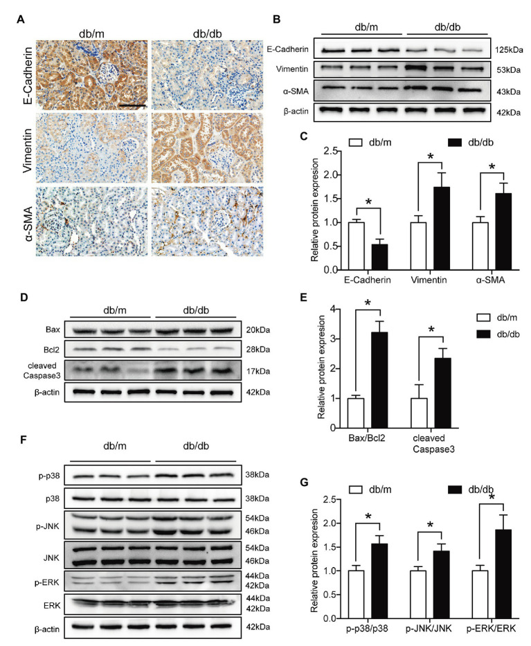
Figure 7.
Renal tubular EMT and apoptosis and MAPK pathway activation were exacerbated in type 2 diabetic kidneys. (A) Representative images of immunohistochemical staining of E-cadherin, Vimentin, and α-SMA in kidney sections. Original magnification = 400. Scale bar = 100 μm. (B) Western blot bands showing E-cadherin, Vimentin, and α-SMA protein expression in the kidney samples. (C) Quantitative analysis of (B), N = 9. (D) Western blot bands showing Bax, Bcl2, and cleaved Caspase 3 protein expression in the kidney samples. (E) Quantitative analysis of (D), N = 9. (F) Western blot bands showing p-p38 MAPK, p38 MAPK, p-JNK, JNK, p-ERK, and ERK protein expression in the kidney samples. (G) Quantitative analysis of (F), N = 9. The data are presented as the mean ± SD. * p < 0.05.

