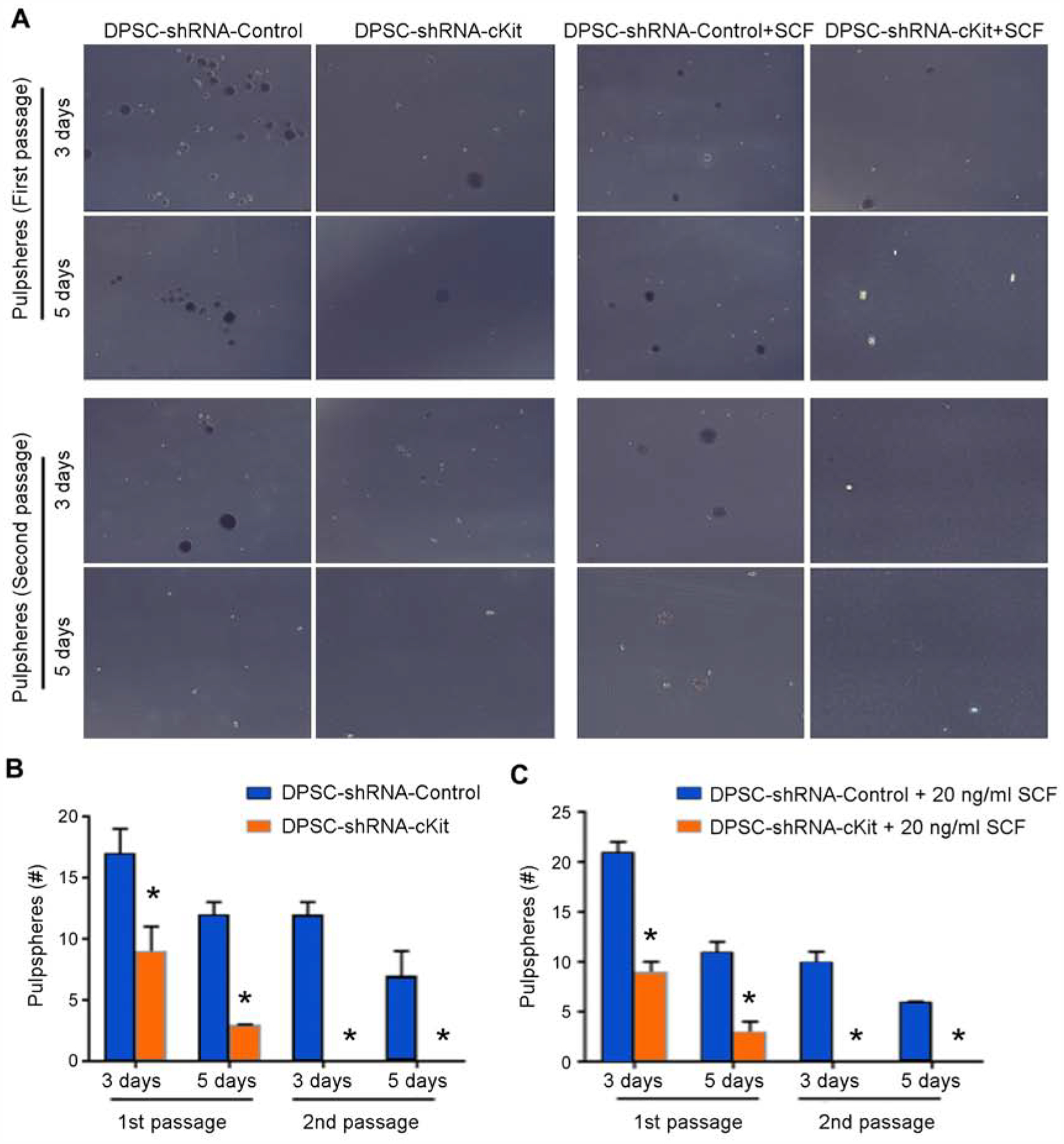
Figure 2:

Dental pulpsphere formation assay. (A) Panel showing DPSC-shRNA-c-Kit and DPSC-shRNA-Control cells in the presence or absence of 20 ng/ml rhSCF in ultra-low attachment plates. (B) Graph showing pulpsphere counts of untreated DPSC-shRNA-c-Kit or DPSC-shRNA-Control cells at days 3 and 5 for both, primary and secondary passage spheres. (C) Graph showing pulpsphere counts of DPSC-shRNA-c-Kit and DPSC-shRNA-Control cells exposed to SCF at days 3 and 5 for both, primary and secondary passage spheres. Asterisks depict statistical significance at p<0.05.
