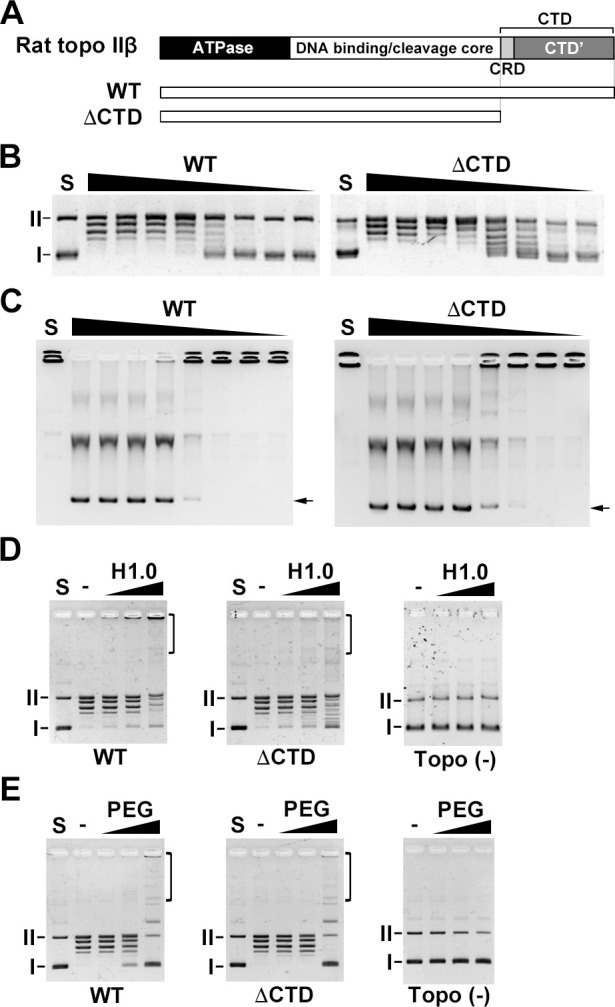
Fig 1. The CTD of rat topo IIβ is required for efficient in vitro catenation.

(A) Structures of rat topo IIβ and the CTD truncation mutant. (B) The relaxation assay was performed with 2-fold serially-diluted enzyme (20 = 100 fmol) and 50 ng pUC18. (C) The decatenation assay was performed with 2-fold serially-diluted enzyme (20 = 100 fmol) and 100 ng kDNA. (D) Purified FLAG-tagged proteins (100 fmol) were used for catenation assay in the presence of histone H1.0 (1, 2 and 4 μg/mL) as a DNA aggregation factor. Deproteinized samples were analyzed by 1% agarose gel. (E) Catenation assay in the presence of PEG (1, 5 and 10%). I: supercoiled DNA. II: nicked circular DNA. Brackets indicate catenanes.
