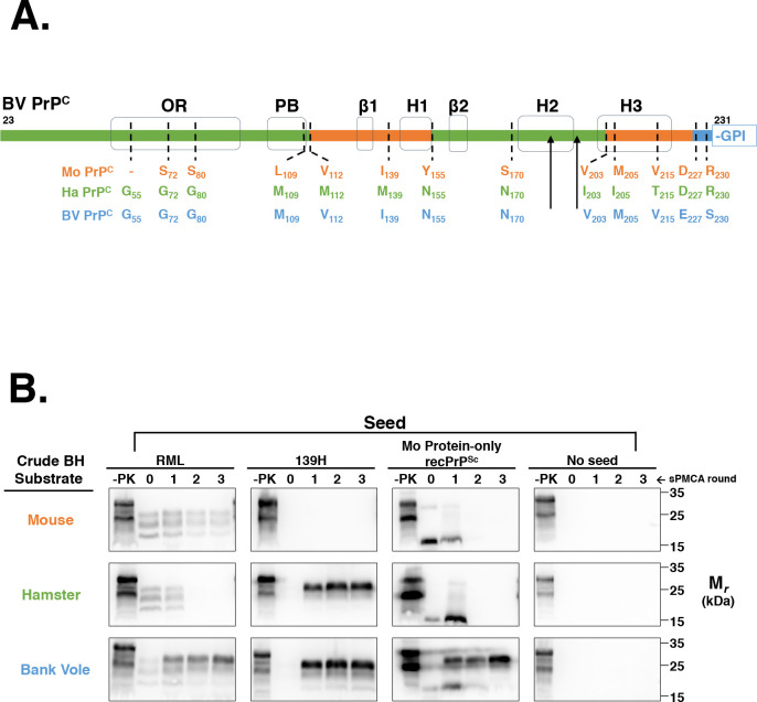
Fig 1. Susceptibility of various rodent species in BH sPMCA.
(A) Amino acid comparison of the processed regions of Mo PrP, Ha PrP, and BV PrP. The sequence bar highlights the regions of BV PrP homologous to Mo PrP (orange) or Ha PrP (green), or are unique to BV PrP (blue). Black arrowheads denote the location of N-linked glycans. The locations of various structural domains are shown in black. OR = octapeptide repeat; PB = polybasic domain; GPI = glycophosphaditylinositol. Sequence alignments were performed using Multalin[25]. (B) Western blots showing three-round sPMCA reactions using normal brain homogenates (BH) from the species used in (A) as the substrates and initially seeded on day 0 with various seeds, as indicated. Day 0 samples are seeded reactions not subject to sonication. -PK = samples not subjected to proteinase K digestion; all other samples were proteolyzed.

