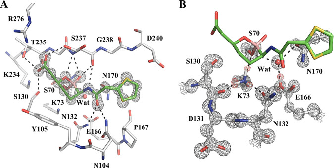
FIG 2.
Vaborbactam complex crystal structure with CTX-M-14 class A β-lactamase. (A) Vaborbactam covalently bound in the active site of CTX-M-14 (1.0-Å resolution; PDB code 6V7H). The unbiased Fo-Fc omit map of vaborbactam is contoured at 3σ and represented with gray mesh. Hydrogen bonds are shown as black dashed lines. (B) Protonation state of K73 and E166 in the CTX-M-14–vaborbactam complex. The 2Fo-Fc omit map (gray) is contoured at 3σ around the protein residues, and the unbiased Fo-Fc omit map (red) is contoured at 2σ around K73 and E166, corresponding to hydrogen atoms located on the terminal groups of K73 and E166.

