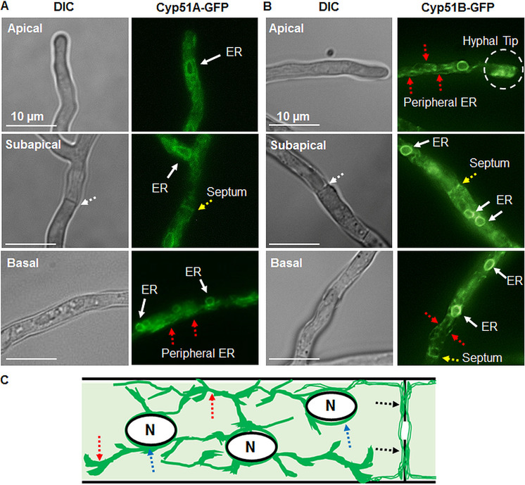
FIG 2.
(A, B) Aspergillus fumigatus Cyp51A and Cyp51B proteins localize to the endoplasmic reticulum (ER) network, including the perinuclear and peripheral ER tubules in the apical, subapical, and basal regions of the hyphae. Red dotted arrows indicate the peripheral ER. The hyphal tip gradient of the ER observed at the hyphal tip is indicated by a dashed circle. Yellow arrows indicate peripheral ER tubules along the septum, and white arrows indicate the perinuclear ER. DIC, differential interference contrast. (C) Pictorial representation of a subapical region showing the Cyp51 protein ER localization patterns in a hyphal compartment. Dotted arrows in blue indicate the perinuclear ER, red dotted arrows indicate the peripheral ER, and black dotted arrows indicate peripheral ER tubular localization along the hyphal septum. N, nucleus.

