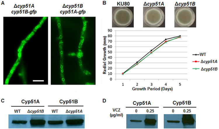
FIG 3.
(A) GFP-tagged Cyp51A or Cyp51B localized in the ER, despite removal of their paralog. (B) Radial growth quantification of the Δcyp51A and Δcyp51B strains in comparison to that of the akuBKU80 (KU80) parent strain. (C) Western blot analysis of the Cyp51A and Cyp51B protein levels in the wild-type (WT), Δcyp51A, and Δcyp51B strains using anti-GFP antibody. (D) Cyp51A and Cyp51B protein expression increased in the presence of voriconazole (VCZ).

