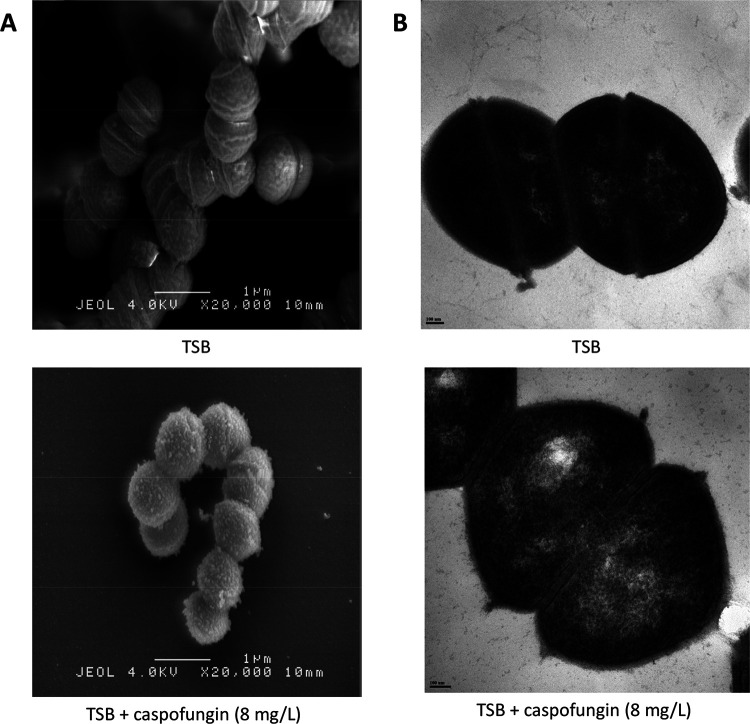
FIG 3.
Cell wall analysis of E. faecium Aus0004 in the presence of caspofungin (8 mg/liter) by electron microscopy. (A) Scanning electron microscopy (SEM) images of E. faecium Aus0004 cells grown in tryptic soy broth (TSB) (top) or TSB plus caspofungin (bottom). Magnification, ×20,000. Morphological abnormalities on cell surface (roughened surface, extrusions) are easily visible. (B) Transmission electron microscopy (TEM) images of E. faecium Aus0004 cells grown in TSB (top) or TSB plus caspofungin (bottom).

